Confirmed speakers

Richard Walton

We lift the suspence and proudly unveil our mystery speaker:
Richard Walton (he/him), University of Bordeaux, France

In vivo multimodal microscopic sub-endocardial imaging using optical catheters in a sheep model of myocardial infarction

There is an unmet capability to identify and localize the combined electrical, structural, or biochemical substrates that predispose to ventricular fibrillation. We aimed to develop of a multimodal imaging fiber probe capable of combined morphological and biochemical characterization of arrhythmogenic substrates at high optical resolutions as a novel cardiac diagnostic tool. A pre-clinical model of chronic myocardial infarction was developed for in vivo investigations. At the chronic phase, structural and compositional characteristics were investigated at the microscopic scale using an optical catheter with integrated electrical mapping. Optical fibres were coupled to a customized multimodal acquisition system incorporating Raman spectroscopy, optical coherence tomography (OCT) and fluorescent imaging. The catheter tip forming the tissue interface was composed of a piezo scanning fibre and lens configuration enabling mapping of the underlying tissue for OCT and autofluorescence imaging. For optimal imaging in the beating heart, optical acquisitions were coordinated with a right ventricle stimulation pulse. After detection of an R wave, a premature stimulation was delivered to interrupt the ventricular capture of the subsequent sinus beat, prolonging the diastolic window absent of cardiac motion. In vivo imaging through the catheter design revealed detailed volumetric images of the endocavity sub-surface microstructure. Healthy myocardium, either surviving myocardial infarction or from sham hearts showed low contrast images of myocardial cell orientation. Conversely, imaging pathological tissue revealed prominent striated patterns of high contrast affiliated with collagen deposition and fibrosis. Concordantly, collagen sheaths surrounding Purkinje fibres were highly visible enabling identification, quantification and morphological analysis of individual Purkinje fibres within free-running and sub-surface branches. We conclude that optical imaging of the microscale in cardiac tissue can be achieved in vivo in the beating heart. Volumetric imaging of heart structure and biochemical composition revealed the morphology and distribution of structurally remodeled tissue. This catheter design offers high potential for multimodal optical applications in cardiac diagnostics and therapy guidance.

Martin Bishop

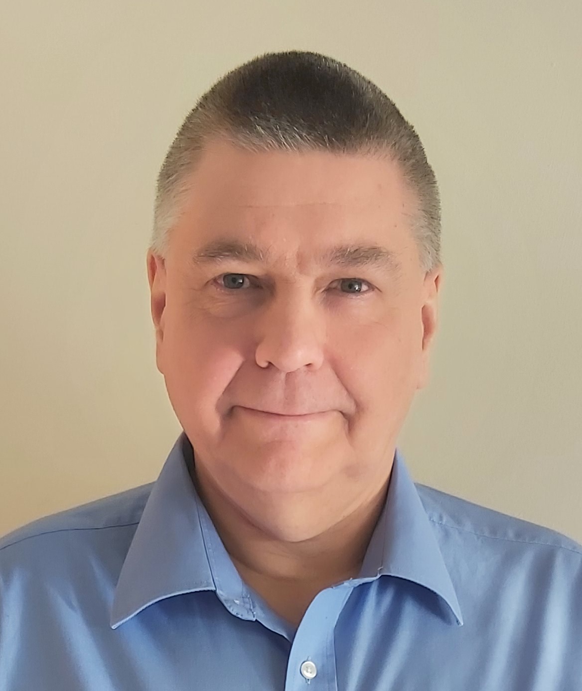
Martin Bishop (he/him), King's College London, UK

Computational simulation in experimental and clinical cardiac electrophysiological research: interactions with novel optical imaging technologies

Computational simulation plays an important role in many different aspects of cardiac electrophysiological research, both in terms of direct clinical translation and also in collaboration with basic science experimentalists, particularly with respect to novel optical imaging technologies. In this short talk, I will begin by briefly discussing how modelling can be used in conjunction with the latest advancements in clinical cardiac imaging to help directly guide ablation therapy, stratify patient risk for lethal arrhythmias and optimise electrotherapy devices and protocols. In the main part of the talk, I will then focus on the utility of modelling in experimental cardiac research, providing recent examples of collaborative projects where modelling has been used to further investigate hypotheses, perform controlled parameter sensitivity analysis and provide high resolution 3-dimensional spatial and temporal data on complex phenomena. I will also show examples of modelling-driven projects which have used novel imaging data for essential validation purposes and those which use the high-resolution structural imaging data as a starting point for highly-detailed model creation. Finally, I will briefly introduce the work we have conducted on simulation of the optical imaging process itself, and how this can be used to facilitate interpretation of experimental imaging data and potentially guide novel clinical optical catheter development.

Tobias Brügmann

Tobias Brügmann (he/him), Institute for Cardiovascular Physiology, University Medical Center Göttingen, Germany

Cardiotoxicity screening with pig ventricular slices

Cardiotoxicity screening is crucial in drug development to reduce risks to patients and economic losses. Different in-vitro approaches have been developed, but they are still lacking normal tissue composition and physiological load. Herein, we explore biomimetic cultivation (BMC) of myocardial slices from healthy pigs as a new cardiotoxicity screening approach. Pig left-ventricular samples were cut into 300 μm thick slices. Slices were pre-stretched by 1.5 mN after installation. The twitch force was recorded during electrical stimulation to determine the force-frequency relationship (FFR), the frequency dependence of contraction duration, the effective refractory period (ERP), and the pacing threshold. Slices generated 2.4 ± 1.4 mN twitch force at 1 Hz electrical pacing and showed a positive FFR, and negative frequency-contraction duration relationship. 300μM Na+ channel blocker lidocaine, increased the pacing threshold by ~300%. The L-type Ca2+ channel activator Bay K8644 (300 nM) increased the twitch force to 295±195% (n=7), while the Ca2+ channel blocker nifedipine (100 nM) decreased the force to 21±5.6% (n=9). The hERG K+ channel blocker dofetilide (3 nM) prolonged the ERP and contraction duration to 220±36% and 161±4% (n=5), respectively. The ß-adrenergic receptor and Ikr inhibitor sotalol (10 μM) prolonged the ERP from ~400 ms to ~600 ms, and addition of 1 μM JNJ 303 further prolonged the ERP to ~850ms, suggesting that the Iks current could be detected after blocking Ikr. Late INa could be provoked by 100 nM dofetilide after 12 h treatment, but not by 100 μM moxifloxacin and were identified by the inhibitory action of 10 μM ranolazine. To validate our new approach, we tested five drug candidates selected from the CiPA list as well as acetylsalicylic acid, PBS, and DMSO as controls in a blinded test and we were able to detect all arrhythmic drugs and their specific effects on cardiac electrophysiology. In conclusion, we established a new approach for cardiotoxicity assessment and demonstrate its efficiency. It can be upscaled to medium throughput screening. Thus, we suggest that our approach can reduce animal numbers and improve the efficiency of preclinical cardiotoxicity screening.

Gil Bub

Gil Bub (he/him), McGill University, Montreal, Canada

High throughput, long duration imaging technologies

Many high-throughput imaging systems use automation to image samples sequentially, which prevents their application to some important experimental targets. For example, rhythms in cultured cardiac tissue evolve over hours or even days, but also display fast transitions between states, necessitating continuous measurement. In these experiments, moving between samples would result in missed data. We introduce a random-access parallel (RAP) imaging modality that uses a novel design inspired by a Newtonian telescope to image multiple spatially separated samples without moving parts or robotics. This scheme enables near-simultaneous image capture of multiple petri dishes and random-access imaging with sub-millisecond switching times at the full resolution of the camera. This enables the RAP system to capture long-duration records from different samples in parallel. The system is demonstrated by continuously imaging multiple cardiac monolayer and Caenorhabditis elegans preparations, effectively in parallel. RAP microscopy, along with several other emerging imaging technologies, enable high-throughput studies of dynamics over a range of time scales.

Francis Burton

Francis Burton (he/him), University of Glasgow, Scotland

Making light work of single cell cardiac electrophysiology: maximising fidelity and throughput

For all studies of cardiac muscle function, the use of fluorescent voltage and calcium sensitive probes is well established, but the limitations of their use are under-appreciated and constrain their use in a number of experimental situations. Furthermore, experimental design frequently requires voltage and calcium to be measured simultaneously at a high acquisition rate (> 100Hz) which puts limitations on the optical setup. An additional useful biomarker is the contraction event initiated by the electrical and subsequent calcium transient. In the context of commercial assay systems, there is the additional need to achieve a high enough throughput to permit 200-400 assays per hour. In this talk, the design of Clyde Bioscience Ltd’s CellOPTIQ™ system and the approach to overcoming specific measurement challenges will be discussed. How parallel measurement could be implemented to further increase throughput will also be considered.

Chris Dunsby

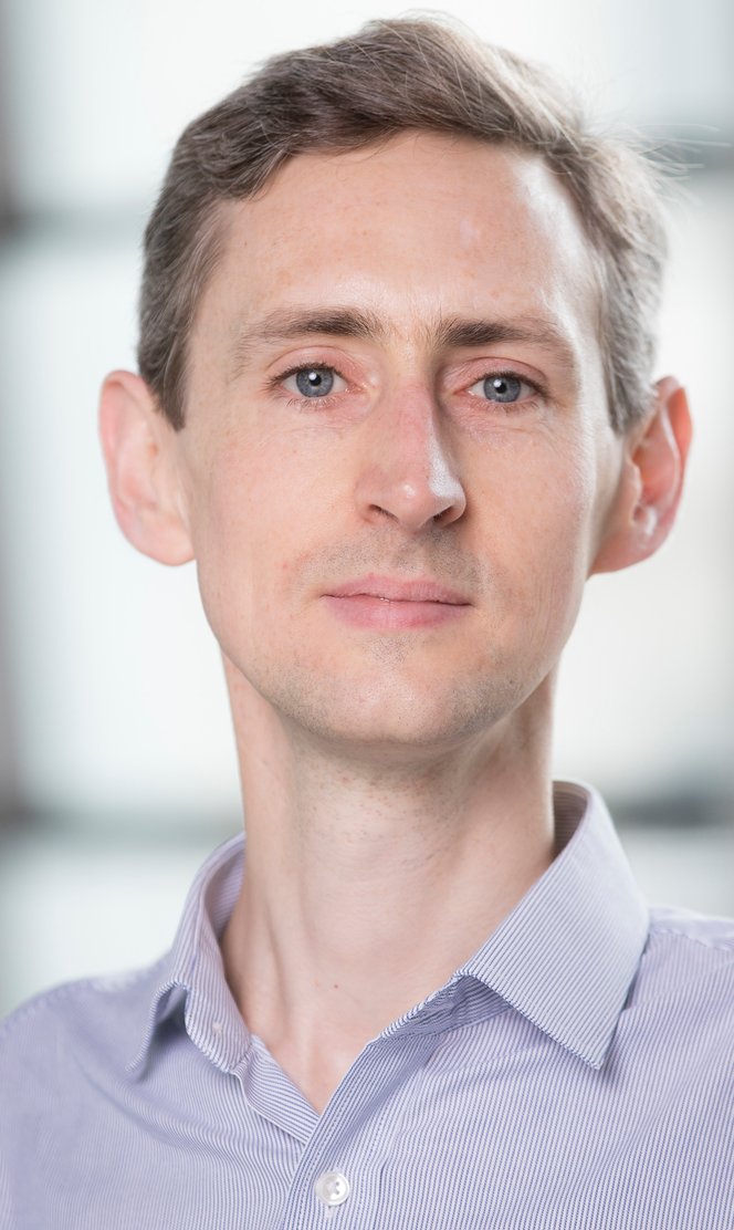
Chris Dunsby (he/him), Imperial College London, UK

High-speed 3D light-sheet microscopy

Light-sheet fluorescence microscopy (LSFM) provides low out-of-plane photobleaching and phototoxicity, but usually requires two microscope objective lenses orientated at 90° to one another – one for fluorescence excitation and one for fluorescence detection – making it harder to image samples prepared using conventional mounting methods. Oblique plane microscopy (OPM) is a type of LSFM that has been developed in my laboratory and uses a single high numerical aperture microscope objective to provide both fluorescence excitation and detection whilst maintaining the advantages of LSFM, enabling it to provide high-speed 3D imaging for a range of applications on a conventional fluorescence microscope frame. The speed of OPM imaging can be applied to image a single sample at video volumetric imaging rates. It can also be used to enable higher throughput and time-lapse 3D imaging of arrays of samples arrayed in multi-well plates. This talk will present examples of the application of OPM for high-speed 3D imaging of isolated cardiomyocytes and also examples where the system is being applied to study arrays of multicellular spheroids and organoids in 3D over multiple conditions and over time.

Sayedeh Hussaini

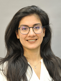
Sayedeh Hussaini (she/her), Max Planck Institute for Dynamics and Self-Organisation, Göttingen, Germany

Temporal modulation of cardiac excitability using optogenetics: exceptional efficacy in terminating cardiac arrhythmia

Introduction: Optogenetics enables remote monitoring and cell-specific control of genetically modified biological systems, such as the heart, by illumination. Over the past decade, cardiac optogenetics has been explored from basic to translational research, particularly in the area of control of cardiac arrhythmia. A better mechanistic understanding of the onset, progression, and control of cardiac arrhythmias benefits the development of alternative methods to conventional treatments, which are often associated with significant side effects for patients. To this end, as numerous studies have found, optogenetics is a promising tool for these fundamental research. Methods: We study the control of arrhythmias in N=5 intact Langendorff-perfused murine hearts expressing ChR2 using two protocols: (i) a single light pulse (SP) (duration 10 and 100 ms, wavelength 470 nm) and (ii) resonant feedback pacing (RFP) with a sequence of global light pulses (duration 20 ms, wavelength 470 nm). The termination success rate is determined as a function of light intensity (LI) for both protocols. ECG recording and potentiometric optical mapping (dye Di-4-ANBDQPQ) are used to measure cardiac activation before, during, and after optical control. Corresponding numerical simulations of cardiac tissue are performed using the Bondarenko model coupled with a channelrhodopsin-2 model in a 2D domain 25x25 mm2 . Results: For the RFP method, the dose-response curve shows a termination rate of more than 50% at the lowest LI of 3.1 μW/mm2, while the SP method achieves this rate at a LI two orders of magnitude higher (LI50SP = 150 or 300 μW/mm2). Moreover, the termination rate at a LI of 100 μW/mm2 is 100% and 45% for the two control methods of RFP and SP, respectively. Numerical simulations show a dose-response consistent with the experimental findings. At very low LIs, simulations suggest that the underlying mechanism for arrhythmia termination is the spatial shift of the reentry core, termed drift, caused by temporal modulation of cardiac tissue excitability during resonant feedback pacing. Conclusion: Using optogenetics we show that resonant feedback pacing has an exceptional efficiency in terminating arrhythmias in numerical simulations and experimentally in intact mouse hearts.

Izzy Jayasinghe

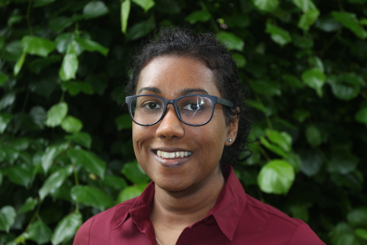
Izzy Jayasinghe (she/her), School of Biosciences, The University of Sheffield, UK

Correlative super-resolution microscopy for understanding the structural basis of fast, calcium signals in the heart

Super-resolution microscopy (SRM) and molecule counting algorithms have advanced our understanding of the clustering properties of membrane proteins like the giant ryanodine receptor (RyR). The positions of RyR channels resolved with these methods have been key enablers for in silico simulations of the intracellular calcium (Ca2+) signals (e.g. Ca2+ sparks, transients or waves) produced by RyRs. In the presented work, we use a recent correlative imaging protocol [1] that experimentally correlates sub-sarcolemmal spontaneous Ca2+ sparks in right ventricular myocytes in rats, recorded with total internal reflection fluorescence (TIRF) microscopy, with the underlying sub-sarcolemmal RyR patterns, visualised with the SRM techniques, DNA- point accumulation for imaging in nanoscale topography (DNA-PAINT). Local examination of the RyR patterns underneath each Ca2+ spark revealed a steep correlation between the size of the Ca2+ sparks and the underlying channel ensembles which typically ranged from ~ 5 to 80 channels. A weaker correlation was observed in right ventricular myocytes isolated from rats with right ventricular failure, reflecting dysfunction in the local regulation of the RyRs in such pathologies. A non-random patterns of Ca2+ sparks featured ‘hot spots’ of distinct sub-cellular regions consisting of recurring spontaneous sparks. Overlay of these regions of high spontaneous activity revealed sub-micron scale groups of 4-8 RyR clusters in healthy right ventricular myocytes. Myocytes studied from failing right ventricular myocytes revealed mixed population of larger RyR clusters and highly fragmented (smaller) clusters within the ‘hot spots’. We conclude that the structure-function correlative imaging offers an approach to locally examine the structural remodelling of RyR clusters that accompany changes in fast cytoplasmic Ca2+ signalling in cardiac pathology.

Daniël Pijnappels

Daniël Pijnappels (he/him), Laboratory of Experimental Cardiology, Department of Cardiology, Leiden University Medical Center, Netherlands

Optical control of bioelectricity to explore biological defibrillation

The implantable cardioverter defibrillator (ICD) is the product of electronic engineering and contains metal, software, and wires to establish a system in which both a detector (i.e. sensor of electrical activity) and effector (i.e. high voltage electroshock generator) are incorporated. Nature also makes use of such detector–effector systems where they control different physiological processes. Such systems are the product of cells, genes, and proteins and involve a regulated variable, set point, and signalling between detector and effector. Given their highly conservative nature, these biological systems are considered to be effective means to rapidly respond to sudden changes that, if sustained, may cause harm. Blood flow and immune regulation are examples of such systems. In contrast, the heart appears to be devoid of a robust biological detector–effector system for counteracting sustained hazardous arrhythmias. The heart does however, have many different ion channels, which generate various electrical currents by opening and closing. This so-called gating is mainly voltage-dependent, meaning that certain ion channels in the cardiomyocyte membrane only open and close upon sensing a certain voltage. An ion channel can therefore be considered as a functional detector–effector unit. This notion creates a rationale to engineer an ion channel protein and integrate it into the plasma membranes of cardiomyocytes to serve the same function as an ICD device, namely rapid detection and termination of arrhythmias. Such fully biological defibrillation would not only have the benefit of being devoid of any hardware, but would also be pain-free given its mode of arrhythmia termination.

Leonardo Sacconi

Leonardo Sacconi (he/him), INO-CNR, Florence, Italy

Correlating electrical disfunction and structural remodeling in Arrhythmogenic Mouse Hearts by advanced optical methods

Severe remodeling processes may occur in the heart due to both genetic and non-genetic diseases. Structural remodeling, such as collagen deposition (fibrosis) and cellular misalignment, can affect electrical conduction at different orders of magnitude and, eventually, lead to arrhythmias. In this scenario, arrhythmogenic cardiomyopathy (ACM) is an inherited heart disease that involves ventricular dysfunction, arrhythmias and localized replacement of contractile fibers with fibrofatty scar tissue. Unfortunately, nowadays, predicting the impact of fine structural alterations on the electrical disfunction in entire organs is challenging, due to the inefficacy of standard imaging methods in performing high-resolution three-dimensional reconstructions in massive tissues. In this work, we developed a new full-optical correlative approach to quantify and integrate the electrical dysfunctions with three-dimensional structural reconstructions of entire hearts, both in controls and in a mouse model of ACM. We combined optical mapping of the action potential propagation (APP) with advances in tissue clearing and light-sheet microscopy techniques. First, we employed an optical platform to map and analyze the APP in Langendorff-perfused hearts. Then, we optimized the SHIELD procedure for the clearing of cardiac tissue, thus converting the previously electrically characterized samples into well-preserved and fully-transparent specimens. A high-throughput light-sheet microscope has been developed following the mesoSPIM project: the conceived microscope allows the reconstruction of the whole mouse heart with micrometric resolution allowing fine quantification of myocytes alignment and fibrosis deposition across the organ. Finally, we developed a software pipeline that employs high-resolution 3D images to analyze and co-register APP maps with the 3D anatomy, contractile fibers disarray, and fibrosis deposition on each heart. We believe that this promising methodological framework will allow clarifying the involvement of fine structural alterations in the electrical dysfunctions, thus enabling a unified investigation of the structural causes that lead to electrical and mechanical alterations after the tissue remodeling.

Franziska Schneider-Warme

Franziska Schneider-Warme (she/her), University Heart Centre Freiburg - Bad Krozingen and Medical Faculty, University of Freiburg, Germany

Optogenetic manipulation of the heterocellular heart

The electromechanical activity of the heart is essential for continuous, coordinated pumping of blood via the circulatory system. The heart itself is a complex multi-cellular organ, consisting of cardiomyocytes (CM) and non-myocytes (NM), the latter including interstitial cells, resident immune cells, and intracardiac neurons, among others. While CM drive the electromechanical function of the heart, cardiac NM have diverse roles, including structural support, signalling and regulatory functions, and maintenance of tissue homeostasis. Following acute cardiac injury such as myocardial infarction, NM proliferate, invade from the circulation, and follow defined activation programmes, thus acting as important players in the process of cardiac scar formation. We assess principles of CM↔NM communication in healthy and diseased myocardium. More specifically, we study electrophysiological interactions between cardiac NM, specifically fibroblasts (FB) and tissue-resident macrophages (MΦ), and CM, with a focus on hitherto unproven FB→CM and CM→MΦ crosstalk. To this end, we combine newly developed optogenetic approaches with state-of-the-art structural (tissue clearing, super-resolution fluorescence microscopy), electrophysiological (patch-clamp, sharp electrode recordings, optical mapping of membrane voltage dynamics), and computational methods. Our study provides a quantitative assessment of structural remodeling and re-distribution of FB, MΦ and collagen in situ in post-injury hearts. Furthermore, data from optogenetic experiments indicate that channelrhodopsin-mediated depolarization of NM can alter electrical conduction by modulating action potential duration and restitution in the scar and scar border zone.

Jonathan Taylor

Jonathan Taylor (he/him), School of Physics and Astronomy, University of Glasgow, Scotland

Optical-computational techniques for timelapse imaging of heart structure and function in vivo.

The heart is a particularly challenging organ to image in 3D timelapse, due to its constant motion. To image processes on timescales of minutes to hours (such as heart development, cell migration, repair and regeneration) demands some form of synchronized image acquisition in order to isolate the high-frequency heartbeat motion from the lower-speed morphological changes of interest. It is also important that any solution should avoid undesirable physiological effects on the sample. Our techniques for prospective optically-gated light sheet microscopy have given us a routine tool for day-long 3D timelapse imaging of the developing zebrafish heart, enabling us to image immune cell dynamics during the inflammatory response to cardiac injury, and revealing unexpected proliferative behaviour of cardiomyocytes during trabeculation. We have also been able to map out blood flow fields within the beating heart, by using related synchronization techniques to extract high-precision measurements from noisy and low-quality raw image data. Not only does this work offer a bridge between direct experimental measurements and computational fluid-structure modelling of heart development, it also points the way towards exciting possibilities in integrated studies of the developmental coupling between heart structure, fluid flow and electrophysiology. Studies like these highlight the fact that “resolution” in a microscope does not tell the whole story. We can only get the most out of the microscope if we are able to cope with motion artefacts, and can find ways to extract high-quality measurements from noisy data, and establish these techniques as routine tools for biological research labs.

Elen Tolstik

Elen Tolstik (she/her), Leibniz-Institut für Analytische Wissenschaften – ISAS – e.V., Germany

Raman spectroscopy of the heart

Nondestructive and label-free Raman imaging has already demonstrated its applicability in life sciences, especially in cancer, neurodegenerative and cardiovascular diseases, with potential clinical applications. From healthy to sick stages, the tissue changes are very small and required the inclusion of extensive data analysis to identify them. Molecular characterization of heart biopsies is well accomplished using Raman-based approaches. These include confocal Raman microspectroscopy, coherent anti-Stokes Raman scattering (CARS) microscopy, surface-enhanced spectroscopy (SERS), and several more. The contrast obtained in imaging cardiac tissue or myocardial cells is based on the vibrational frequencies of functional groups, which provide us with specific molecular "fingerprints" and corresponding structural information of the probes under investigation. This presentation will review recent applications of vibrational spectroscopy in lysosomal storage diseases with strong cardiac involvement. It will be shown how disease-associated biomolecular changes in the heart can be detected with high sensitivity, thus evaluating the potential of Raman spectroscopy for early diagnosis of metabolic and storage diseases.